
California Optomap
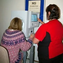
Optos’ widefield retinal imaging technology has the potentialto offer your eyecare physician the most powerful tools for disease diagnosis and management. Theoptomap images provide enhanced clinical information which facilitates the early detection, management and treatment of disorders and diseases evidenced in the retina such as retinal detachments and tears, glaucoma, diabetic retinopathy and age-related macular degeneration. Retinal imaging can also indicate evidence of non-eye or systemic diseases such as hypertension and certain cancers.

Nerve Fiber Analyzer
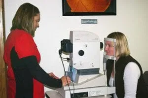
Optical Coherence Tomography (OCT) is a non-invasive technology used for imaging the retina, the multi-layered sensory tissue lining the back of the eye. OCT, the first instrument to allow doctors to see cross-sectional images of the retina, is revolutionizing the early detection and treatment of eye conditions such as macular holes, pre-retinal membranes, macular swelling and even optic nerve damage. OCT testing has become a standard of care for the assessment and treatment of most retinal conditions. OCT uses rays of light to measure retinal thickness and can be performed in a few minutes. No radiation or x-rays are used in this test.
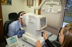
Topography Corneal Topography is non-invasive medical imaging technique for mapping the surface curvature of the cornea. Since the corena is normally responsible for some 70% of the eye's refractive power, its topology is of critical importance in determing your quality of vision. The three-dimensional map is a valuable aid to the examination process and can assist in the diagnosis and treatment of a number of conditions; in planning refractive surgery such as LASIK and evaluation of its results; or in assessing the fit of contact lenses. The procedure is carried out in seconds and is completely painless.
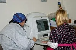
Retinal Photography The digital retinal imaging camera takes an image of the retina (the back of the eye). These pictures are necessary to document the health of the optic nerve, vitreous, macula, retina and its blood vessels. The photographs are used for comparison, documentation, and sometimes to diagnose certain eye conditions. This is helpful in obtaining a baseline for future care and monitoring active diseases such as macular degeneration and glaucoma. A picture is worth a thousand words!
Visual Fields The visual fields instrument provides information regarding how well the optic nerve and retina are functioning. It is useful in diagnosing and following certain conditions such as glaucoma.